Lectin Array

10605

From: $527.00

ZBiotech has developed a robust microarray platform that allows researchers to simultaneously profile glycosylation by multiple lectins. The lectin array utilizes a panel of 40 plant-based lectins for high-throughput glycosylation analysis. Each lectin has been validated by glycan microarrays to ensure binding specificity. The proprietary coating substrate of this array outperforms other substrates with exceptional sensitivity without compromising specificity. Each lectin array contains 8 or 16 identical subarrays, enabling the analysis of multiple samples simultaneously and facilitating rapid results in a simple and easy-to-use format. Varieties of biological samples can be analyzed by the array. For example, proteins, antibodies, cells, cell lysate, serum, vesicles, bacteria, and viral particles. The Lectin array can be customized to meet individual client needs. Assay services are available upon request.

SKU: 10605 Category:
Description
Structures
Examples
Citations
Document

Description

Glycosylation is one of the most abundant posttranslational modifications in eukaryotic cells. It is a complex process in which multiple glycosyltransferases work together to covalently add carbohydrate structures to proteins, lipids, or ribonucleic acids. Glycosylation is an intracellular process essential to all eukaryotic cells. The carbohydrate structures stabilize proteins to ensure proper folding for optimal activities. They also involve cellular signaling regulation to enforce tissue homeostasis. Determining the chemistry and biology of glycosylation is indispensable for understanding the biology of living organisms.

The discovery of ricin by Stillmark in 1888 marked the beginning of lectinology, a discipline dedicated to studying lectins’ chemical and biological functions. In 1952, Watkins and Morgan found that adding sugars can block the agglutination of erythrocytes caused by lectins. This milestone discovery unveiled the carbohydrate-binding specificity of lectins. Since then, hundreds of lectins have been purified and studied from plants, microorganisms, and animals. Lectins are now defined as carbohydrate-binding proteins, and each lectin recognizes a unique repertoire of carbohydrate structures.

For example, SNA (Sambucus Nigra lectin), MAL1 (Maackia Amurensis Lectin I), and MAL2 (Maackia Amurensis Lectin II) are sialoglycan-specific lectins. SNA recognizes terminal α-2,6 sialic acids (either Neu5Ac or Neu5Gc). In stark contrast, MAL1 and MAL2 preferentially recognize terminal α-2,3 sialic acids (either Neu5Ac or Neu5Gc). Another example is Calsepa (Calystegia Sepium lectin). Calsepa generally binds with all N-linked glycans without differentiating a specific subtype of N-glycans with or without terminal sialic acids. Compared with Calsepa, BanLec (Musa Paradisiaca lectin) preferentially recognizes high-mannose N-glycans and hybrid N-glycans without terminal sialic acids.

Lectins have been widely used to detect glycosylation because of their unique carbohydrate-recognizing properties. ZBiotech has developed a robust microarray platform that allows researchers to simultaneously profile glycosylation by multiple lectins. The lectin array utilizes a panel of 40 plant-based lectins immobilized on the array surface for high-throughput glycosylation analysis. Each lectin has been validated by glycan microarrays to ensure binding specificity. The proprietary coating substrate of this array outperforms other substrates with exceptional sensitivity without compromising specificity. Each lectin array contains 8 or 16 identical subarrays, enabling the analysis of multiple samples simultaneously and facilitating rapid results in a simple and easy-to-use format. Varieties of biological samples can be analyzed by the array. For example, proteins, antibodies, cells, cell lysate, serum, vesicles, bacteria, and viral particles. The Lectin array can be customized to meet individual client needs. Assay services are available upon request.

Features

  • 40 lectins (validated by glycan microarrays)
  • Unrivaled sensitivity and specificity;
  • Simple assay format;
  • Small sample volume;
  • Customizable (select lectins for a specific microarray format)
  • Assay service available;

Applications

  • Evaluate glycosylation of proteins;
  • Evaluate glycosylation of antibodies;
  • Evaluate glycosylation of viral particles;
  • Evaluate glycosylation of bacteria;
  • Evaluate glycosylation of cells;
  • Evaluate glycosylation of vesicles;

Structures

List of lectins and their binding specificities (download the PDF)

ID Lectin Binding Specificity
NL1 AAL Fucose-containing glycans; no binding to fucose on Blood Group A and B antigens except minor binding to type 2 antigens.
NL2 AIA O-glycans containing following motifs: T antigen (Core 1); Tn antigen; O-GalNAc Core 3; no binding to STn, Core 2, Core 4 and Core 6 O-GalNAc glycans, Blood Group ABH Antigens Type 3.
NL3 BC2L-C Fucose; no or weak binding to Blood Group A and B type 2 antigens.
NL4 BPL Terminal β-Gal/β-GalNAc: PolyLacNAc > Mono/Di LacNAc. Sialic acid attached to Gal shields the binding. Fucose attached to GlcNAc allows the binding; no binding to Blood Group Antigens.
NL5 Calsepa Bind to N-glycans; no difference between bisecting N-glycans, complex N-glycans and core-fucose N-glycans.
NL6 ConA High-mannose, complex, hybrid, bisecting N-glycans; no binding to tri- and tetra-antennary N-glycans.
NL7 DBA Glycans with Sda antigen motif (e.g., GM2); Blood Group A antigen.
NL8 DSA Type 2 LacNAc; (GlcNAcβ1-4)2 and some extended epitopes; bind to Blood Group antigen A, B, H, Type 2.
NL9 ECL Terminal β1,4-Gal; no binding to Lewis X glycan; bind to Blood Group Antigen H Type 2.
NL10 GNA Exposed α1,3-Man on N-glycans
NL11 GSI-B4 Terminal α1,3-Gal (or weaker α1,4-Gal); bind to Blood Group B Antigens.
NL12 GSII Terminal GlcNAc
NL13 HAA Terminal α-GalNAc: Blood Group A > GalNAca1-3GalNAcb > All other α-GalNAc; Similar to HPA.
NL14 HHL High Mannose N-glycans; Weak binding to Complex (non-bisecting GlcNAc) N-Glycans and other mannose polysaccharides; no binding to bisecting N-glycans.
NL15 HPA Terminal GalNAc; weak binding to T antigen; no binding to Sda antigen, Blood Group A Antigen. Similar to HAA.
NL16 LCA Core-fucose N-glycans; High-mannose N-glycans.
NL17 LEL LacNAc including Blood Group ABH antigens; weak binding to High-mannose N-glycans; α2,3-sialylated type 2 poly-LacNAc glycans.
NL18 LTA Terminal Fucα1-3GlcNAc; Lewis X; Lewis Y; Blood Group H disaccharide, Type 2 and 6.
NL19 MAL-I Terminal Neu5Ac(or Neu5Gc)α2-3Galβ1-4GlcNAc(or Glc), or 3′-sulfated LacNAc or Lactose; weak binding to asialo-glycans; no binding to SLeX or Sda antigen.
NL20 MAL-II Terminal Neu5Ac(or Neu5Gc)α2-3Galβ1-4GlcNAc(or Glc), or 3′-sulfated LacNAc or Lactose; weak binding to asialo-glycans; no binding to SLeX or Sda antigen.
NL21 Morniga G High-mannose N-glycan > Poly-LacNAc N-glycan > Tri-antennary N-glycans
NL22 NPA High-mannose N-glycans > Hybrid N-glycans > Core-fucose N-glycans
NL23 PHA-E Complex N-Glycans; no high-mannose binding
NL24 PHA-L Bisecting N-glycans
NL25 PNA Galβ1-3GalNAc (T antigen)
NL26 PSA Core N-Glycan Fucose (Fuca1-6GlcNAc). No binding to non-fucosylated N-Glycans.
NL27 PTA Bind to Blood Group H antigens except Type 1.
NL28 RCA-I Terminal β1,4-Gal (β1,3 and β1,6 also accepted);  no binding with terminal α2,3 sialic acid, or to T antigen or Lewis X; still binds to β1,4-Gal capped with terminal α2,6 sialic acid or 6′-sulfate.
NL29 RPA Complex N-Glycans: Tetra-antennary > Tri-antennary; weak binding to Poly-LacNAc extended bi-antennary; no Other bi-antennary N-Glycan binding.
NL30 RSL Fucose-containing glycans; includes all blood group fucose, core N-Glycan fucose ,and Lewis A/X Fucose
NL31 SBA Terminal α-GalNAc or β-GalNAc; Tn antigen; MUC-1 glycopeptides; similar to VVL
NL32 SNA Terminal Neu5Ac(or Neu5Gc)α2-6Galβ1-4; no asialo partial-epitope binding.
NL33 STL Galβ1-4GlcNAc (type 2 poly-LacNAc) with weak partial-epitope binding; weak binding to High-mannose N-Glycans (GlcNAcB1-4GlcNAc epitope); bind to Blood Group ABH antigens type 2.
NL34 UEA-I Fucα1-2Galβ1-4GlcNAc (Blood Group H Type 2, 5, 6, disaccharide); Lewis Y;  α1,4-Fuc
NL35 VVL Tn antigen; weak binding to blood group antigen A
NL36 WFA Terminal GalNAc; terminal β-Gal on LacNAc; no binding to Lewis X. bind to Sda antigen; no T antigen binding.
NL37 WGA GlcNAc; terminal and exposed α2,3-Neu5Ac; terminal and exposed α-GalNAc;  no binding to Lewis X;  no binding with terminal α2,6 sialic acid.
NL38 RPL-Sia2 Neu5Acα2-3Galβ1-3GalNAc glycans (e.g. ST antigen)
NL39 RPL-Man2 High-mannose or Hybrid N-glycans
NL40 RPL-αGal Terminal α-Gal glycans

Examples

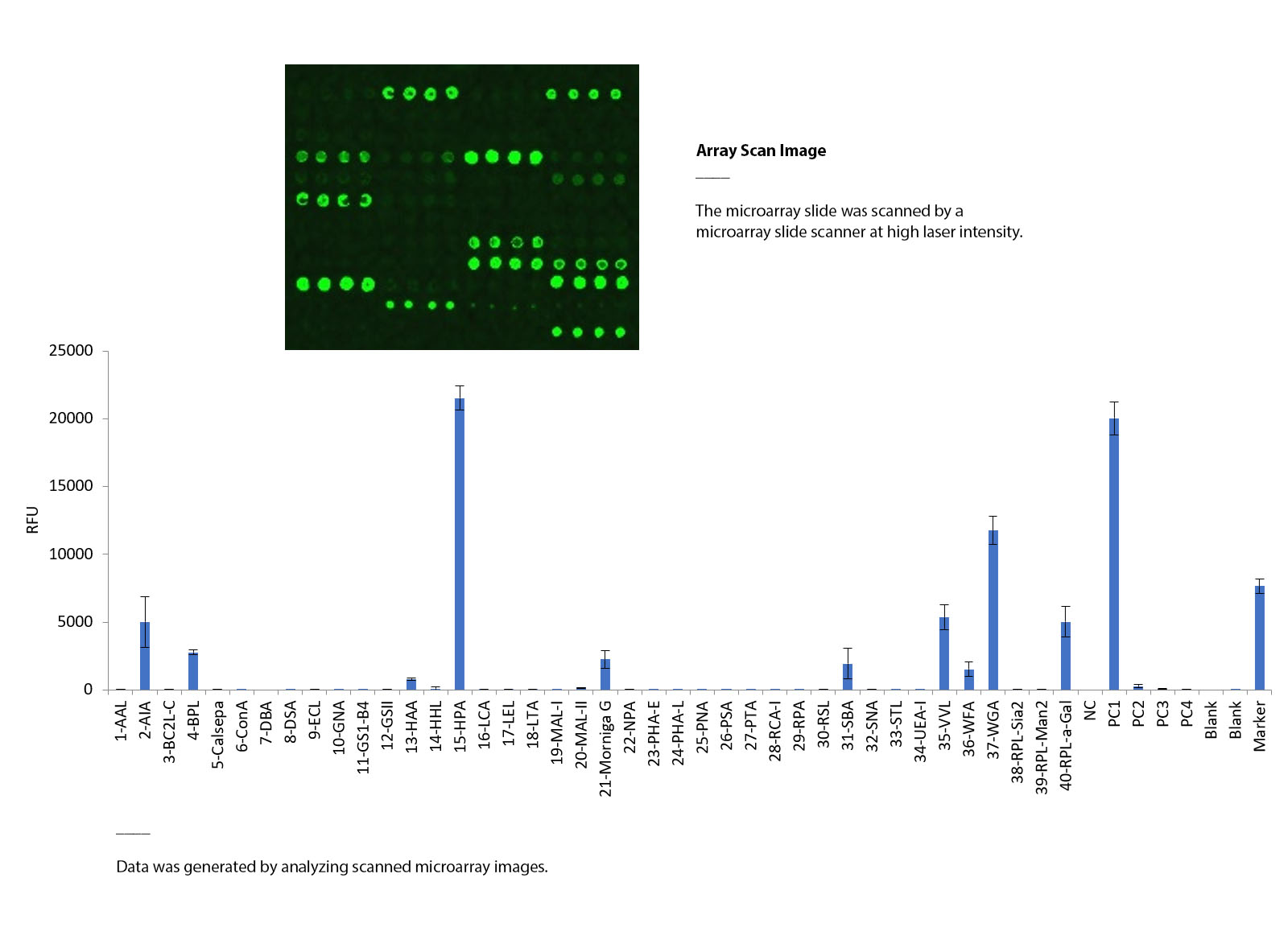
Assessing the binding specificity of α-GalNAc-PAA through lectin array analysis

To assess the binding specificity of α-GalNAc-PAA, a subarray of the lectin microarray slide was assayed with a biotinylated α-GalNAc-PAA at a concentration of 5 μg/ml, followed by detection with streptavidin-Cy3. The microarray slide was scanned using an InnoScan 710 microarray scanner at 2 PMT and low laser power at a 532nm wavelength. Negative control spots showed no non-specific binding, while positive control 1 and the marker displayed expected signals. Notably, lectins such as HPA and VVL that have known binding specificity to α-GalNAc glycans also showed expected binding signals in the analysis. These results provide valuable insights into the binding specificity of α-GalNAc-PAA and demonstrate the utility of lectin microarray analysis for detecting the presence of glycan epitopes.

Document

List of lectins and their binding specificities (download the PDF)

Protocol & User Manual (download the manual)