From: $994.00
The Sulfated Glycan Array provides researchers with a powerful tool to study the interactions between sulfated glycans and a wide range of biological samples, such as proteins, antibodies, cell lysates, serum, vesicles, bacteria, or viral particles. This innovative array features 46 structurally-defined glycans, including paired sulfated glycans and their non-sulfated counterparts. The glycan repertoire includes diverse types such as O-glycans, O-mannose glycans, and various small glycan motifs, offering a comprehensive platform for sulfated glycan-binding studies.
Each array is equipped with 8 or 16 identical subarrays, enabling the simultaneous analysis of multiple samples. Results can be generated within hours, facilitating quick and efficient exploration of sulfated glycan interactions. Assay services are available upon request to support your experimental goals.
Sulfated glycans are a distinct class of sugar molecules characterized by the addition of sulfate groups to their structures. These modifications play crucial roles in a variety of biological processes, including cellular communication, immune response, and the regulation of signaling pathways. Sulfated glycans are especially known for their ability to mediate interactions with proteins, such as lectins and growth factors, influencing key physiological and pathological processes. For instance, sulfated glycans have been implicated in modulating inflammation, inhibiting viral entry, and regulating tumor progression.
The biological activities of sulfated glycans are often linked to their specific structural motifs, which include the arrangement of monosaccharides, linkage types, and sulfate group positions. These motifs determine the binding affinity and specificity of sulfated glycans toward various biological targets. Due to their importance, sulfated glycans have been investigated as biomarkers for diseases and as therapeutic targets for drug development. Moreover, paired comparisons of sulfated glycans with their non-sulfated forms provide valuable insights into the functional roles of sulfation in glycan activity.
Innovative Surface Chemistry for Enhanced Multivalent Display of Sulfated Glycans
The multivalent presentation of sulfated glycans is especially important for biomolecule recognition due to the inherent negative charge associated with these glycans. This cumulative negative charge within a cluster of sulfated glycans significantly influences biomolecule recognition processes.
To achieve efficient multivalent presentation of sulfated glycans on the microarray surface, we have developed a novel approach by modifying the conventional planar NHS coating chemistry. This novel approach involves introducing proprietary highly-branched spherical polymers along with a specialized linker, which serves to bridge the polymer and NHS functional group. Unlike the traditional planar NHS coating, where one substrate unit carries only one NHS functional group, our innovative multivalent NHS coating allows for variable numbers of NHS functional groups per substrate unit. This unique spherical polymer enables the presentation of multiple glycans per substrate, facilitating effective multivalent presentation.
Features
Applications
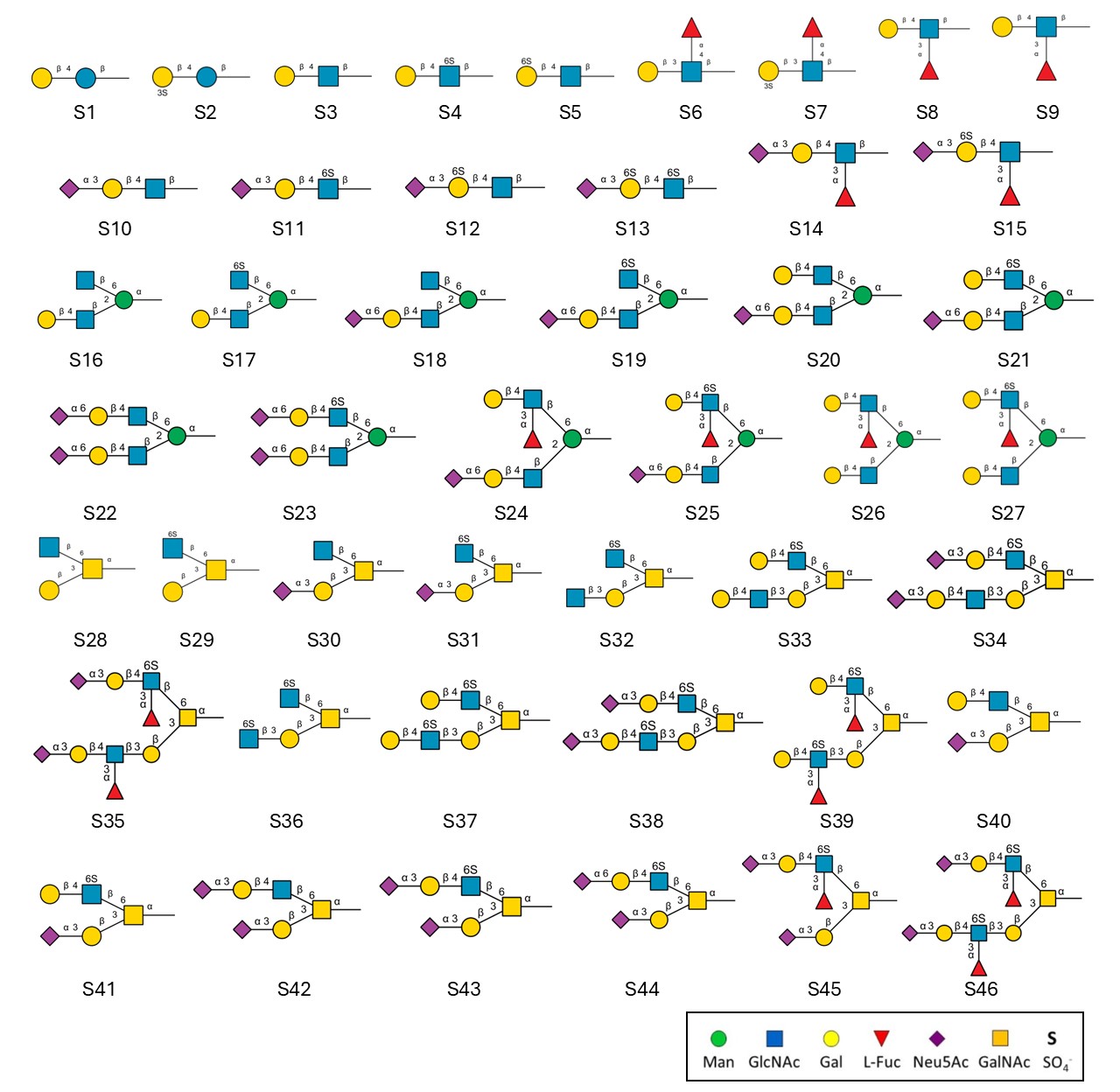
List of Sulfated glycan structures on the array (download the PDF)
ID | Structure |
---|---|
S1 | Galβ1-4Glcβ |
S2 | (3S)Galβ1-4Glcβ |
S3 | Galβ1-4GlcNAcβ |
S4 | Galβ1-4(6S)GlcNAcβ |
S5 | (6S)Galβ1-4GlcNAcβ |
S6 | Galβ1-3(Fucα1-4)GlcNAcβ |
S7 | (3S)Galβ1-3(Fucα1-4)GlcNAcβ |
S8 | Galβ1-4(Fucα1-3)GlcNAcβ |
S9 | (3S)Galβ1-4(Fucα1-3)GlcNAcβ |
S10 | Neu5Acα2-3Galβ1-4GlcNAcβ |
S11 | Neu5Acα2-3Galβ1-4(6S)GlcNAcβ |
S12 | Neu5Acα2-3(6S)Galβ1-4GlcNAcβ |
S13 | Neu5Acα2-3(6S)Galβ1-4(6S)GlcNAcβ |
S14 | Neu5Acα2-3Galβ1-4(Fucα1-3)GlcNAcβ |
S15 | Neu5Acα2-3(6S)Galβ1-4(Fucα1-3)GlcNAc |
S16 | GlcNAcβ1-6(Galβ1-4GlcNAcβ1-2)Manα |
S17 | (6S)GlcNAcβ1-6(Galβ1-4GlcNAcβ1-2)Manα |
S18 | GlcNAcβ1-6(Neu5Acα2-6Galβ1-4GlcNAcβ1-2)Manα |
S19 | (6S)GlcNAcβ1-6(Neu5Acα2-6Galβ1-4GlcNAcβ1-2)Manα |
S20 | Galβ1-4GlcNAcβ1-6(Neu5Acα2-6Galβ1-4GlcNAcβ1-2)Manα |
S21 | Galβ1-4(6S)GlcNAcβ1-6(Neu5Acα2-6Galβ1-4GlcNAcβ1-2)Manα |
S22 | Neu5Acα2-6Galβ1-4GlcNAcβ1-6(Neu5Acα2-6Galβ1-4GlcNAcβ1-2)Manα |
S23 | Neu5Acα2-6Galβ1-4(6S)GlcNAcβ1-6(Neu5Acα2-6Galβ1-4GlcNAcβ1-2)Manα |
S24 | Galβ1-4(Fucα1-3)GlcNAcβ1-6(Neu5Acα2-6Galβ1-4GlcNAcβ1-2)Manα |
S25 | Galβ1-4(Fucα1-3)(6S)GlcNAcβ1-6(Neu5Acα2-6Galβ1-4GlcNAcβ1-2)Manα |
S26 | Galβ1-4(Fucα1-3)GlcNAcβ1-6(Galβ1-4GlcNAcβ1-2)Manα |
S27 | Galβ1-4(Fucα1-3)(6S)GlcNAcβ1-6(Galβ1-4GlcNAcβ1-2)Manα |
S28 | GlcNAcβ1-6(Galβ1-3)GalNAcα |
S29 | (6S)GlcNAcβ1-6(Galβ1-3)GalNAcα |
S30 | GlcNAcβ1-6(Neu5Acα2-3Galβ1-3)GalNAcα |
S31 | (6S)GlcNAcβ1-6(Neu5Acα2-3Galβ1-3)GalNAcα |
S32 | (6S)GlcNAcβ1-6(GlcNAcβ1-3Galβ1-3)GalNAcα |
S33 | Galβ1-4(6S)GlcNAcβ1-6(Galβ1-4GlcNAcβ1-3Galβ1-3)GalNAcα |
S34 | Neu5Acα2-3Galβ1-4(6S)GlcNAcβ1-6(Neu5Acα2-3Galβ1-4GlcNAcβ1-3Galβ1-3)GalNAcα |
S35 | Neu5Acα2-3Galβ1-4(Fucα1-3)(6S)GlcNAcβ1-6(Neu5Acα2-3Galβ1-4(Fucα1-3)GlcNAcβ1-3Galβ1-3)GalNAcα |
S36 | (6S)GlcNAcβ1-6((6S)GlcNAcβ1-3Galβ1-3)GalNAcα |
S37 | Galβ1-4(6S)GlcNAcβ1-6(Galβ1-4(6S)GlcNAcβ1-3Galβ1-3)GalNAcα |
S38 | Neu5Acα2-3Galβ1-4(6S)GlcNAcβ1-6(Neu5Acα2-3Galβ1-4(6S)GlcNAcβ1-3Galβ1-3)GalNAcα |
S39 | Galβ1-4(Fucα1-3)(6S)GlcNAcβ1-6(Galβ1-4(Fucα1-3)(6S)GlcNAcβ1-3Galβ1-3)GalNAcα |
S40 | Galβ1-4GlcNAcβ1-6(Neu5Acα2-3Galβ1-3)GalNAcα |
S41 | Galβ1-4(6S)GlcNAcβ1-6(Neu5Acα2-3Galβ1-3)GalNAcα |
S42 | Neu5Acα2-3Galβ1-4GlcNAcβ1-6(Neu5Acα2-3Galβ1-3)GalNAcα |
S43 | Neu5Acα2-3Galβ1-4(6S)GlcNAcβ1-6(Neu5Acα2-3Galβ1-3)GalNAcα |
S44 | Neu5Acα2-6Galβ1-4(6S)GlcNAcβ1-6(Neu5Acα2-3Galβ1-3)GalNAcα |
S45 | Neu5Acα2-3Galβ1-4(Fucα1-3)(6S)GlcNAcβ1-6(Neu5Acα2-3Galβ1-3)GalNAcα |
S46 | Neu5Acα2-3Galβ1-4(Fucα1-3)(6S)GlcNAcβ1-6(Neu5Acα2-3Galβ1-4(Fucα1-3)(6S)GlcNAcβ1-3Galβ1-3)GalNAcα |
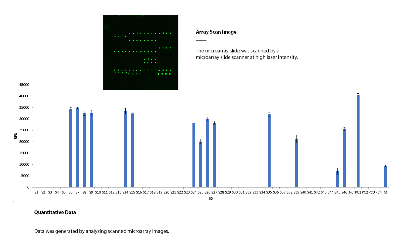
Aleuria aurantia lectin (AAL) binds to various sulfated and non-sulfated glycans that contain fucose.
The Sulfated Glycan Array was tested using biotinylated AAL lectin (5 μg/mL), followed by streptavidin-Cy3 (0.2 μg/mL). The array was scanned with a microarray scanner at a wavelength of 532 nm, and the positive controls demonstrated binding signals as expected. AAL exhibited binding to multiple sulfated and non-sulfated glycans with fucose present.
Human Siglec-8 and Its Interaction with Sulfated Glycans
Human Siglec-8, a sialic acid-binding immunoglobulin-like lectin, is predominantly expressed on eosinophils and mast cells and plays a significant role in regulating immune responses. It is known to bind selectively to specific glycans, particularly sulfated glycans such as 6′-sulfo-sLex, which contribute to its functional specificity in various physiological and pathological processes, including inflammation and allergy regulation.
The Sulfated Glycan Array was used to investigate the binding interactions of human Siglec-8. The assay utilized the extracellular binding domain of human Siglec-8 fused to an human IgG Fc tag (2.5 μg/mL), followed by detection with anti-human IgG Fc (2.5 μg/mL). Scanning at a wavelength of 532 nm revealed binding signals. Human Siglec-8 exhibited binding to multiple sulfated glycans.
List of Sulfated glycan structures on the array (download the PDF)
Protocol & User Manual (download the manual)