From: $599.00
The Pan-Gal and GalNAc Detection Kit offers a precise and innovative solution for detecting terminal Galactose (Gal) and N-Acetylgalactosamine (GalNAc) residues on glycans. The kit leverages Galactose oxidase to specifically oxidize terminal Gal and GalNAc, creating aldehyde groups that are uniquely reactive. These aldehydes are then labeled through aniline-catalyzed oxime ligation with biotin tags. The biotinylated glycans can be detected using fluorescently labeled streptavidin, enabling compatibility with flow cytometry analysis. Compared to traditional lectin-based approaches, which may have limited coverage or specificity, this method ensures a broader and more accurate detection of general terminal Gal and GalNAc residues across diverse glycoconjugates. The kit includes all essential reagents for performing up to 20 tests, along with a step-by-step protocol to ensure ease of use and reproducibility. For added flexibility, we also provide related assay and analysis services tailored to your experimental requirements.
Galactose oxidase (GAO) is an oxidoreductase enzyme that specifically catalyzes the oxidation of primary alcohol groups in galactose and its derivatives to form aldehydes. This enzyme plays a pivotal role in various biochemical applications due to its substrate specificity and efficiency in generating reactive aldehydes under mild conditions. The Pan-Gal and GalNAc Detection Kit leverages GAO to enable the detection of terminal Galactose (Gal) and N-Acetylgalactosamine (GalNAc) residues, offering researchers a tool for studying glycan modifications.
The Mechanism Behind Pan-Gal and GalNAc Detection
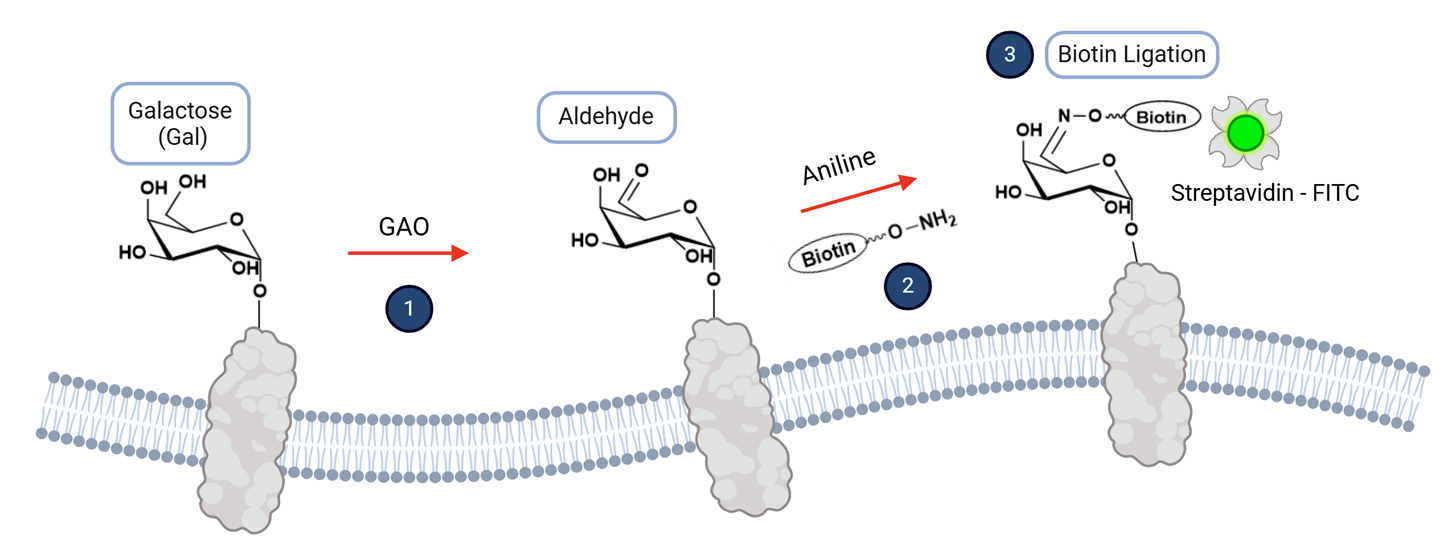
The Pan-Gal and GalNAc Detection Kit utilizes a robust and specific enzymatic-chemical method to detect terminal Gal and GalNAc residues on glycans (Figure 1):
Figure 1. Illustration of Terminal Gal/GalNAc Labeling with Biotin for Flow Cytometry Analysis
The first step involves the enzymatic oxidation of terminal Gal and GalNAc residues using galactose oxidase. GAO targets the specific hydroxyl groups of these sugars, converting them into reactive aldehyde groups. This step is highly specific, ensuring minimal interference from other glycan components, and operates under mild conditions to preserve the integrity of the sample.
Once the aldehyde groups are generated, they undergo aniline-catalyzed oxime ligation with aminooxy-biotin. Aniline serves as a nucleophilic catalyst, significantly enhancing the efficiency of the oxime bond formation. This step ensures the stable and effective biotin tagging of the oxidized Gal and GalNAc residues, making them detectable in subsequent assays. The oxime ligation is both rapid and highly specific, providing reliable labeling results.
The biotinylated glycans are detected using fluorescently labeled streptavidin, which binds with high affinity to biotin. For flow cytometry analysis, a fluorescent label, such as FITC, is conjugated to streptavidin, enabling the visualization and quantification of the labeled glycans. The fluorescence intensity corresponds to the levels of terminal Gal and GalNAc residues, providing researchers with a quantitative measure of these glycans on live cells.
This kit offers a streamlined workflow with all necessary reagents for performing up to 20 tests, ensuring reproducibility and ease of use. The innovative enzymatic and chemical approach sets it apart from traditional lectin-based methods, delivering superior coverage and specificity in the detection of terminal Gal and GalNAc residues. For added flexibility, related assay and analysis services are available to tailor the solution to your research needs.
Assay Kit Specifications
The Pan-Gal and GalNAc Detection Kit includes all the necessary reagents for performing up to 20 tests. Each kit contains:
For optimal results, we recommend using 4.0-6.0 x 10^5 cells per analysis. This cell range ensures sufficient detection and accurate analysis of terminal Gal and GalNAc residues. To determine the optimal concentration of galactose oxidase for different cell or sample types, we suggest performing a concentration optimization experiment. This involves testing 3-5 recommended concentrations of galactose oxidase to oxidize the samples and determine the saturation point. This step is crucial since the levels of terminal Gal and GalNAc residues can vary among different samples, and identifying the best concentration will maximize the efficiency and accuracy of the assay.
Customization and Services
In addition to the assay kit, we offer customized assay services tailored to meet specific experimental requirements, allowing researchers to leverage our expertise for their unique research needs.
Oxidation Levels and Terminal Gal/GalNAc Labeling
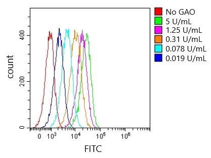
Human embryonic kidney (HEK293) cells were treated with galactose oxidase at various concentrations to achieve different levels of oxidation. Following oxidation, the cells were ligated with biotin using an aniline-catalyzed oxime ligation protocol and analyzed for terminal Gal and GalNAc labeling. The results demonstrate that varying the concentration of galactose oxidase correlates with the degree of biotin labeling, reflecting different levels of terminal Gal and GalNAc residues (Figure 1).
Figure 1. Concentrations of GAO affect the oxidation levels, which directly correlate with the levels of biotin labeling on the terminal Gal/GalNAc.
Impact of Galactose Oxidase on Terminal Sialic Acids
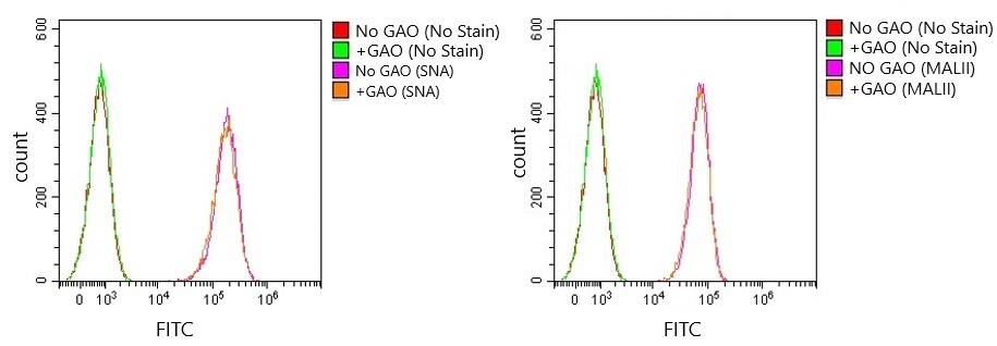
To validate whether galactose oxidase (GAO) oxidation affects terminal sialic acids, HEK293 cells treated with GAO were analyzed using two lectins, SNA and MALII, which specifically recognize terminal sialic acids with α2,6- and α2,3-linkages, respectively. Following GAO treatment, no significant changes in SNA or MALII binding were observed, indicating that GAO oxidation does not impact terminal sialic acids. This result confirms the specificity of GAO for oxidizing terminal Gal and GalNAc residues without interfering with sialic acid structures.
Figure 2. GAO oxidation does not affect Terminal Sialic Acids
Impact of Galactose Oxidase on Terminal Galactose
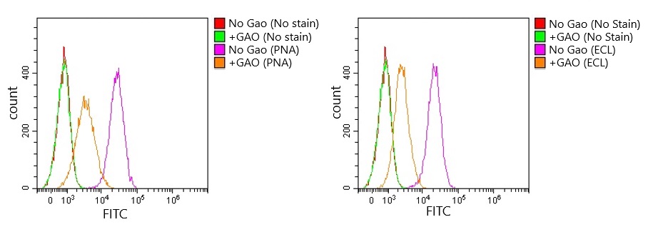
To validate whether galactose oxidase (GAO) oxidation disrupts terminal Gal residues, HEK293 cells were treated with GAO and subsequently analyzed using two lectins, PNA and ECL, which specifically recognize terminal Gal. Following GAO treatment, a significant reduction in PNA and ECL binding was observed. This indicates that GAO effectively oxidizes terminal Gal residues, disrupting their structure as expected. These findings confirm that GAO targets and modifies terminal Gal, validating its functional specificity for this sugar residue.
Figure 3. GAO oxidation alters Terminal Galactose
Neuraminidase Treatment Increases Exposure of Gal/GalNAc
HEK293 cells were treated with either a control buffer or neuraminidase from Clostridium perfringens (400 mU/mL) to remove terminal sialic acids. Following treatment, the cells were analyzed using the Pan-Gal/GalNAc Detection Kit, with galactose oxidase (GAO) applied at a concentration of 10 U/mL. The removal of sialic acids was confirmed using two lectins, SNA and MALII, which specifically recognize terminal sialic acids. Neuraminidase treatment significantly reduced binding to both SNA and MALII, validating the removal of terminal sialic acids.
Moreover, the neuraminidase treatment increased the exposure of terminal Gal and GalNAc residues on the cell surface. This was reflected by the higher levels of biotin-labeled Gal/GalNAc detected after neuraminidase treatment, indicating that the enzymatic removal of sialic acids unmasked additional Gal and GalNAc residues.
Figure 4. Neuraminidase treatment reduces SNA and MALII binding, confirming the removal of sialic acids from the cell surface, and increases the exposure of GAO-oxidized Gal/GalNAc residues.