Alpha 2,3/2,6 Sialylation Profiling Kit

10901-30T-1

From: $599.00

The Alpha 2,3/2,6 Sialylation Profiling Kit enables researchers to analyze specific alpha 2,3 and alpha 2,6 sialic acids on live mammalian cells using flow cytometry. This specialized kit includes six validated lectins for precise detection and differentiation of these sialic acids. These lectins include alpha 2,3 sialic acid-preferred or specific lectins such as MALII, RPL-Sia1, and RPL-Sia2, and alpha 2,6 sialic acid-preferred lectin such as SNA, along with two lectins that recognize non-sialylated glycans: ECL, which recognizes terminal beta 1,4-Gal, and PNA, which recognizes T antigen. The kit provides a complete set of reagents necessary for the assay. Additionally, we offer an assay service to perform the analysis, tailored to meet your specific experimental needs.

SKU: 10901-30T-1 Category:
Description
Examples

Description

Sialylation, the addition of sialic acid residues to glycans, plays essential roles in various biological processes, including cell-cell interaction, immune response, and pathogen recognition. There are different linkages of sialic acids, notably alpha 2,3 and alpha 2,6, which have distinct biological implications and associations with various diseases. Alpha 2,3-linked sialic acids are often involved in cancer progression and metastasis. For example, increased levels of alpha 2,3 sialylation have been linked to the invasive potential of certain cancer cells. On the other hand, alpha 2,6-linked sialic acids are critical in immune regulation and have been implicated in autoimmune diseases. Differentiating between these sialic acid linkages is crucial for understanding disease mechanisms.

Assay Kit and Lectin Specificity

The Alpha 2,3/2,6 Sialylation Profiling Kit provides a comprehensive set of six widely used lectins to analyze the presence of alpha 2,3 and alpha 2,6 sialic acids on live cells. Each lectin in this kit has a specific binding preference, enabling detailed profiling of sialylation patterns:

  • SNA (Sambucus nigra agglutinin): Preferentially recognizes alpha 2,6 sialic acids decorated on certain glycans.
  • MALII (Maackia amurensis lectin II): Preferentially recognizes alpha 2,3 sialic acids decorated on certain glycans.
  • RPL-Sia1 (Recombinant Prokaryotic Lectins): Binds to terminal alpha 2,3-linked sialic acid (Neu5Ac) on both N-linked and O-linked glycans.
  • RPL-Sia2 (Recombinant Prokaryotic Lectins): Specific for terminal alpha 2,3-linked sialic acid (Neu5Ac) on O-linked glycans.
  • PNA (Peanut agglutinin): Recognizes T antigen, an O-glycan without terminal sialic acid.
  • ECL (Erythrina cristagalli lectin): Recognizes terminal beta 1,4-Gal without terminal sialic acid.

Kit Contents and Specifications

This assay kit is sufficient for 20 tests for each lectin and includes the following components:

  • Biotinylated SNA
  • Biotinylated MALII
  • Biotinylated RPL-Sia1
  • Biotinylated RPL-Sia2
  • Biotinylated PNA
  • Biotinylated ECL
  • FITC-conjugated streptavidin for detection
  • Relevant buffers

For optimal results, we recommend using 2.0-5.0 x 10^5 cells per analysis. To ensure accurate and reliable results, we recommend performing an optimization experiment for each lectin on your specific cell types. Since the presence of sialic acids can vary among different cell lines, and using excessive lectin can trigger cell death, it is crucial to identify the optimal cell number and lectin concentration. This step will help achieve consistent and reproducible outcomes.

Customized Services

In addition to the assay kit, we offer customized services to perform the analysis, tailored to meet your specific experimental needs, leveraging our expertise for your unique research applications.

Examples

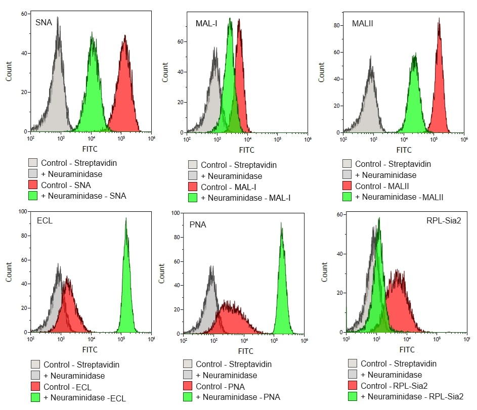
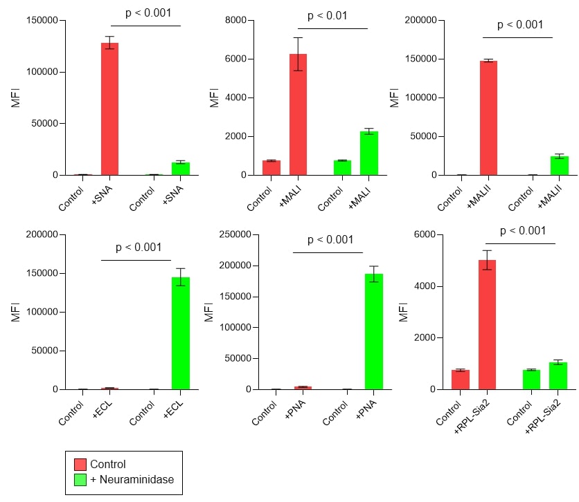
Human melanoma cells (A375) were treated with either a control solution or neuraminidase from Clostridium perfringens (400 mU/mL). The treated cells were then stained with the indicated biotinylated lectins and detected with streptavidin-FITC. The data indicate that neuraminidase treatment significantly reduced SNA, MALII, RPL-Sia1, and RPL-Sia2 bindings, while it enhanced PNA and ECL bindings as terminal non-sialic acids were exposed. This data suggests that the panel of lectins works as intended, demonstrating the specificity and effectiveness of the assay kit.

Figure 1. Representative overlapping histogram for each lectin analyzed.


Figure 2. Quantification of mean fluorescence intensity (MFI) for each lectin analyzed.