Glycosphingolipid Glycan Array

10608

From: $1,222.00

Glycosphingolipid (GSL) glycan microarray technology is developed to allow researchers to explore the interactions between GSL glycans and biological samples such as proteins, antibodies, cells, cell lysate, serum, vesicles, bacteria, or viral particles. The GSL glycan array features 58 distinct GSL glycan structures. Each array contains 8 or 16 identical subarrays, enabling the simultaneous analysis of multiple samples. The GSL glycan array provides high-throughput and reliable glycan-binding information with a simple assay format that only requires a small sample volume. It can be customized to meet individual client needs. Assay services are available upon request.

SKU: 10608 Category:
Description
Structures
Examples
Document

Description

Glycosphingolipids (GSLs), a subclass of glycolipids, are central components of the cellular membrane. A GSL molecule comprises a hydrophobic ceramide backbone covalently linked to a hydrophilic glycan moiety. The glycan moieties are either neutral, sialylated, or sulfated. Glycan portions of GSLs can be further classified based on the sequence and configuration of the sugar moieties into globo-, lacto-, neoLacto-, and ganglioside types of GSL glycans. These glycan structures are linked to various ceramides, creating structurally diversified GSLs.

In normal physiological conditions, GSL glycans play diverse roles in maintaining membrane structure, transducing cellular signals, and providing scaffolds for cell-to-cell interactions. However, abnormal presentations of these glycans have been linked to metabolic diseases, cancer, and neurodegenerative diseases. For example, the aberrant expression of specific GSL glycans is a hallmark of cancer cells, altering a range of cellular functions to promote survival. Deregulation of GSL glycans also impacts neuroinflammation associated with Parkinson’s. Therefore, identifying and understanding the differences between GSL glycans expressed in normal and pathological conditions is vital for understanding the pathogenesis of these diseases.

GSL glycans have gained increasing interest in the biomedical research community. ZBiotech has developed a robust microarray platform that allows researchers to explore the interactions between GSL glycans and biological samples, such as proteins, antibodies, cells, cell lysate, serum, vesicles, bacteria, or viral particles. The GSL glycan array features 58 artificially synthesized GSL glycan epitopes immobilized on a solid array surface for binding analysis. It provides useful glycan-binding information for evaluating GSLs – based biological interactions. Each array contains 8 or 16 identical subarrays, enabling the simultaneous analysis of multiple samples. The GSL Glycan Array provides high-throughput and reliable glycan-binding information with a simple assay format that only requires a small sample volume. It can be customized to meet individual client needs. Assay services are available upon request.

Features

  • Unrivaled sensitivity and specificity;
  • Simple assay format;
  • Small sample volume;
  • Customizable (select GSL glycans for a specific microarray format)
  • Assay service available;

Applications

  • Evaluate binding specificities of GSL glycan-interacting proteins;
  • Evaluate binding specificities of GSL glycan-interacting antibodies;
  • Study virus – GSL glycan interactions;
  • Study bacteria – GSL glycan interactions;
  • Study vesicle – GSL glycan interactions;
  • Study cell – GSL glycan interactions;

Structures

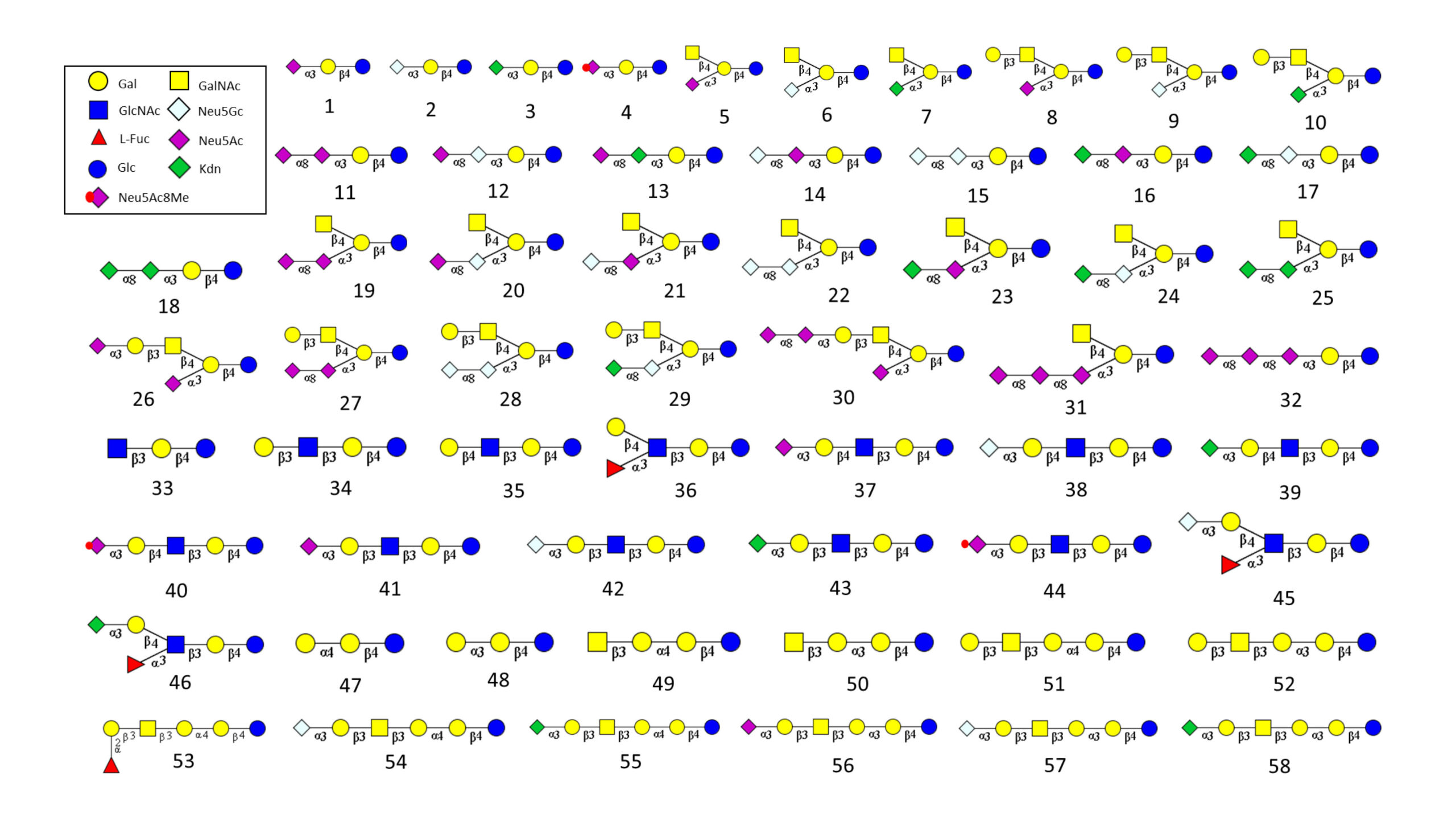
List of GSL glycan structures on the array (download the PDF)

Type ID Glycan Structure Abbreviation
Ganglioside G1 Neu5Acα2–3Galβ1–4Glc Ac-GM3
G2 Neu5Gcα2–3Galβ1–4Glc Gc-GM3
G3 Kdnα2–3Galβ1–4Glc Kdn-GM3
G4 Neu5Ac8Meα2–3Galβ1–4Glc Ac8Me-GM3
G5 Neu5Acα2–3(GalNAcβ1–4)Galβ1–4Glc Ac-GM2
G6 Neu5Gcα2–3(GalNAcβ1–4)Galβ1–4Glc Gc-GM2
G7 Kdnα2–3(GalNAcβ1–4)Galβ1–4Glc Kdn-GM2
G8 Neu5Acα2–3(Galβ1–3GalNAcβ1–4)Galβ1–4Glc Ac-GM1
G9 Neu5Gcα2–3(Galβ1–3GalNAcβ1–4)Galβ1–4Glc Gc-GM1
G10 Kdnα2–3(Galβ1–3GalNAcβ1–4)Galβ1–4Glc Kdn-GM1
G11 Neu5Acα2–8Neu5Acα2–3Galβ1–4Glc Ac-Ac-GD3
G12 Neu5Acα2–8Neu5Gcα2–3Galβ1–4Glc Ac-Gc-GD3
G13 Neu5Acα2–8Kdncα2–3Galβ1–4Glc Ac-Kdn-GD3
G14 Neu5Gcα2–8Neu5Acα2–3Galβ1–4Glc Gc-Ac-GD3
G15 Neu5Gcα2–8Neu5Gcα2–3Galβ1–4Glc Gc-Gc-GD3
G16 Kdnα2–8Neu5Acα2–3Galβ1–4Glc Kdn-Ac-GD3
G17 Kdnα2–8Neu5Gcα2–3Galβ1–4Glc Kdn-Gc-GD3
G18 Kdnα2–8Kdnα2–3Galβ1–4Glc Kdn-Kdn-GD3
G19 Neu5Acα2–8Neu5Acα2–3(GalNAcβ1–4)Galβ1–4Glc Ac-Ac-GD2
G20 Neu5Acα2–8Neu5Gcα2–3(GalNAcβ1–4)Galβ1–4Glc Ac-Gc-GD2
G21 Neu5Gcα2–8Neu5Acα2–3(GalNAcβ1–4)Galβ1–4Glc Gc-Ac-GD2
G22 Neu5Gcα2–8Neu5Gcα2–3(GalNAcβ1–4)Galβ1–4Glc Gc-Gc-GD2
G23 Kdnα2–8Neu5Acα2–3(GalNAcβ1–4)Galβ1–4Glc Kdn-Ac-GD2
G24 Kdnα2–8Neu5Gcα2–3(GalNAcβ1–4)Galβ1–4Glc Kdn-Gc-GD2
G25 Kdnα2–8Kdnα2–3(GalNAcβ1–4)Galβ1–4Glc Kdn-Kdn-GD2
G26 Neu5Acα2–3Galβ1–3GalNAcb1–4(Neu5Aca2–3)Galβ1–4Glc Ac-Ac-GD1a
G27 Neu5Acα2–8Neu5Acα2–3(Galβ1–3GalNAcβ1–4)Galβ1–4Glc Ac-Ac-GD1b
G28 Neu5Gcα2–8Neu5Gcα2–3(Galβ1–3GalNAcβ1–4)Galβ1–4Glc Gc-Gc-GD1b
G29 Kdnα2–8Neu5Gcα2–3(Galβ1–3GalNAcβ1–4)Galβ1–4Glc Kdn-Gc-GD1b
G30 Neu5Acα2–8Neu5Acα2–3Galβ1–3GalNAcβ1–4(Neu5Aca2–3)Galβ1–4Glc Ac-Ac-Ac-GT1a
G31 GalNAcβ1-4(Neu5Acα2–8Neu5Acα2–8Neu5Acα2–3)Galβ1–4Glc Ac-Ac-Ac-GT2
G32 Neu5Acα2–8Neu5Acα2–8Neu5Acα2–3Galβ1–4Glc Ac-Ac-Ac-GT3
Lacto- and Neolacto-series G33 GlcNAcβ1–3Galβ1–4Glc Lc3
G34 Galβ1–3GlcNAcβ1–3Galβ1–4Glc Lc(LNT)
G35

Galβ1–4GlcNAcβ1–3Galβ1–4Glc

nLc(LNnT)
G36 Galβ1–4(Fucα1–3)GlcNAcβ1–3Galβ1–4Glc Fuc-nLc
G37 Neu5Acα2–3Galβ1–4GlcNAcβ1–3Galβ1–4Glc Ac-nLc
G38 Neu5Gcα2–3Galβ1–4GlcNAcβ1–3Galβ1–4Glc Gc-nLc4
G39 Kdnα2–3Galβ1–4GlcNAcβ1–3Galβ1–4Glc Kdn-nLc4
G40 Neu5Ac8Meα2–3Galβ1–4GlcNAcβ1–3Galβ1–4Glc 8MeAc-nLc4
G41 Neu5Acα2–3Galβ1–3GlcNAcβ1–3Galβ1–4Glc Ac-Lc4
G42 Neu5Gcα2–3Galβ1–3GlcNAcβ1–3Galβ1–4Glc Gc-Lc4
G43 Kdnα2–3Galβ1–3GlcNAcβ1–3Galβ1–4Glc Kdn-Lc4
G44 Neu5Ac8Meα2–3Galβ1–3GlcNAcβ1–3Galβ1–4Glc 8MeAc-Lc4
G45 Neu5Gcα2–3Galβ1–4(Fucα1–3)GlcNAcβ1–3Galβ1–4Glc Gc-Fuc-nLc4
G46 Kdnα2–3Galβ1–4(Fucα1–3)GlcNAcβ1–3Galβ1–4Glc Kdn-Fuc-nLc4
Globo- and Isoglobo-series  G47 Galα1–4Galβ1–4Glc   Gb3
G48 Galα1–3Galβ1–4Glc iGb3
G49 GalNAcβ1–3Galα1–4Galβ1–4Glc  Gb4
G50 GalNAcβ1–3Galα1–3Galβ1–4Glc iGb4
G51 Galβ1–3GalNAcβ1–3Galα1–4Galβ1–4Glc Gb5 (SSEA-3)
G52 Galβ1–3GalNAcβ1–3Galα1–3Galβ1–4Glc iGb5
G53 Fucα1-2Galβ1-3GalNAcβ1-3Galα1-4Galβ1-4Glc Globo-H
G54 Neu5Gcα2–3Galβ1–3GalNAcβ1–3Galα1–4Galβ1–4Glc Gc-Gb5
G55 Kdnα2–3Galβ1–3GalNAcβ1–3Galα1–4Galβ1–4Glc Kdn-Gb5
G56 Neu5Acα2–3Galβ1–3GalNAcβ1–3Galα1–3Galβ1–4Glc Ac-iGb
G57 Neu5Gcα2–3Galβ1–3GalNAcβ1–3Galα1–3Galβ1–4Glc Gc-iGb5
G58 Kdnα2–3Galβ1–3GalNAcβ1–3Galα1–3Galβ1–4Glc Kdn-iGb5

Examples

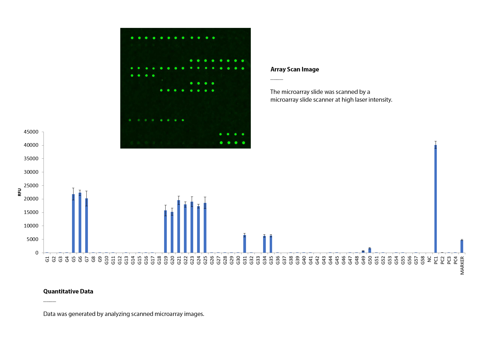
Using GSL glycan microarray to determine the binding specificity of Wisteria Floribunda Lectin (WFA)

The GSL glycan array was assayed with biotinylated Wisteria Floribunda Lectin (10 μg/mL), followed by streptavidin (Cy3). The array was scanned with a microarray scanner at 532nm wavelength. Positive control showed binding signals as expected. WFA binds to a series of GSL glycans (GM2 and GD2).

Document

List of GSL glycan structures on the array (download the PDF)

Protocol & User Manual (download the manual)