From: $994.00
The human milk oligosaccharide (HMO) glycan array allows researchers to explore the interactions between HMO and various biological samples, for example, proteins, antibodies, cells, cell lysate, serum, vesicles, bacteria, or viral particles. The HMO glycan array features 46 structurally – defined oligosaccharides prevalent in human breast milk. Each array slide contains 16 identical subarrays, enabling the simultaneous analysis of multiple samples. Results can be obtained in only a few hours, making investigating HMO binding easy and efficient. The HMO glycan array provides high-throughput and reliable HMO-binding information. It can be customized to meet individual client needs. Assay services are available upon request.
Human milk oligosaccharides (HMOs) are the third most abundant component of human breast milk. They are a class of complex sugar molecules that positively influence infant development. For example, HMOs stimulate the growth of symbiotic bacteria in the gut, protect against infection and disease, and promote the healthy development of the infant’s gut, brain, and immune function. Many of these beneficial effects are associated with specific epitope structures of HMOs. HMOs have been used as biomarkers to evaluate infant digestive development, diarrhea incidence, morbidity and mortality, and risk of developing certain disorders. In addition to their vital roles in early infant development, HMOs have been found to inhibit pathogens and constrain tumor cell growth.
HMOs are structurally diverse, and more than one hundred structurally distinct HMOs have been identified to date. The composition and abundance of HMOs in breast milk vary from woman to woman over lactation. For example, modification by fucosylation is determined by the genetic background of the mother and whether specific fucosyltransferase genes (e.g., FUT2 or FUT3) are present as wild-type, mutated, or deleted. Fucosylation of the HMO backbone further impacts HMO bioactivity. The health benefits of HMOs are increasingly recognized, and enormous studies are underway to understand further their functions, binding partners, and the relationship between their structures and biological roles.
ZBiotech has developed a robust microarray platform that allows researchers to explore the interactions between HMO and various biological samples, for example, proteins, antibodies, cells, cell lysate, serum, vesicles, bacteria, or viral particles. The HMO glycan array features 46 structurally – defined oligosaccharides prevalent in human breast milk. Each array slide contains 16 identical subarrays, enabling the simultaneous analysis of multiple samples. Results can be obtained in only a few hours, making investigating HMO binding easy and efficient. The HMO glycan array provides high-throughput and reliable HMO-binding information. It can be customized to meet individual client needs. Assay services are available upon request.
Features
Applications
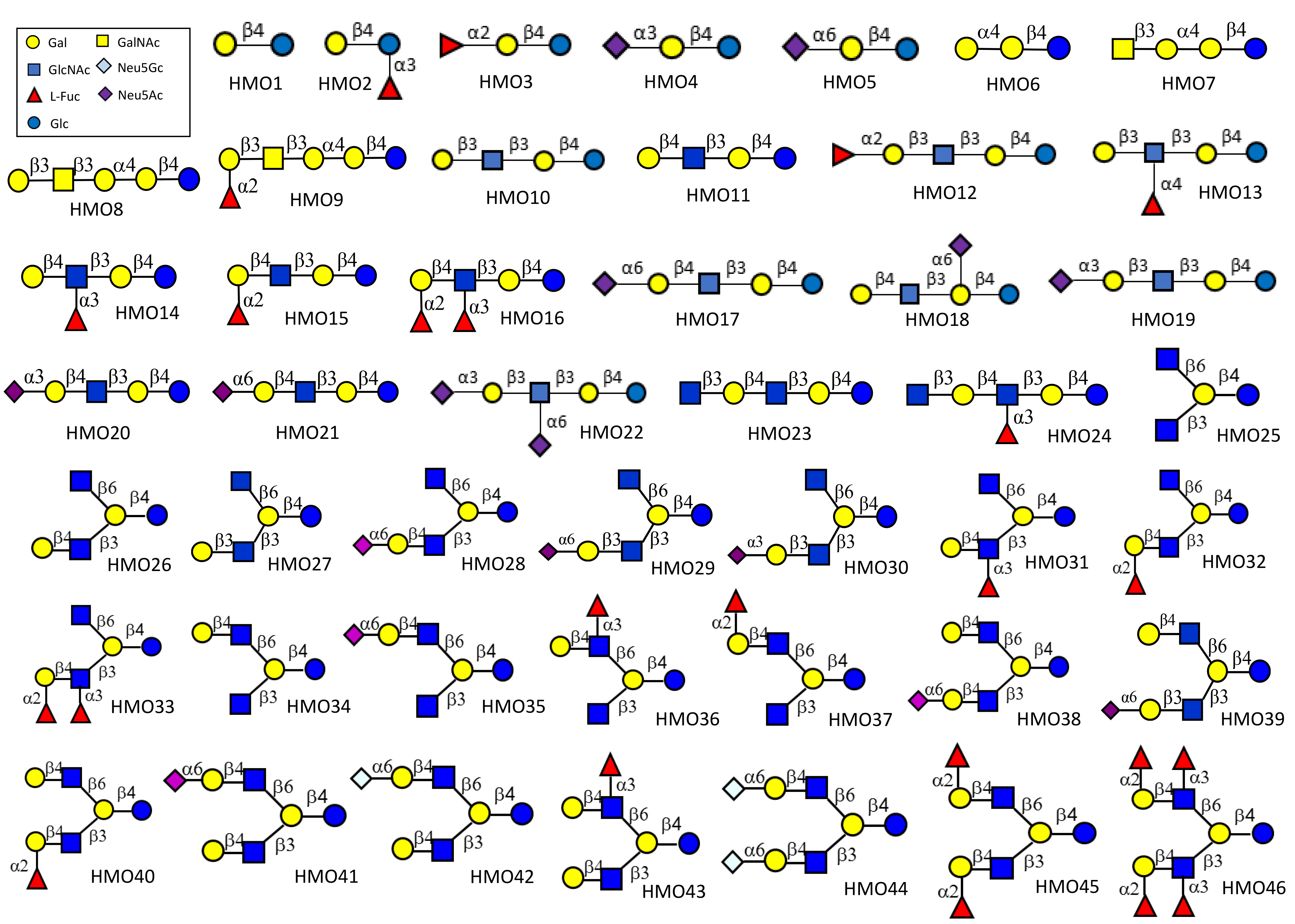
List of human milk oligosaccharide structures on the array (download the PDF)
ID | Structure | Common Name |
---|---|---|
HMO1 | Galβ1-4Glc | Lactose |
HMO2 | Galβ1-4(Fucα1-3)Glc | 3-FL |
HMO3 | Fucα1-2Galβ1-4Glc | 2′-FL |
HMO4 | Neu5Acα2-3Galβ1-4Glc | 3′-SL |
HMO5 | Neu5Acα2-6Galβ1-4Glc | 6′-SL |
HMO6 | Galα1-4Galβ1-4Glc | Gb3 (Pk antigen) |
HMO7 | GalNAcβ1-3Galα1-4Galβ1-4Glc | Gb4 |
HMO8 | Galβ1-3GalNAcβ1-3Galα1-4Galβ1-4Glc | Gb5 (SSEA-3) |
HMO9 | Fucα1-2Galβ1-3GalNAcβ1-3Galα1-4Galβ1-4Glc | Globo-H |
HMO10 | Galβ1-3GlcNAcβ1-3Galβ1-4Glc | LNT |
HMO11 | Galβ1-4GlcNAcβ1-3Galβ1-4Glc | LNnT |
HMO12 | Fucα1-2Galβ1-3GlcNAcβ1-3Galβ1-4Glc | LNFP-I |
HMO13 | Galβ1-3(Fucα1-4)GlcNAcβ1-3Galβ1-4Glc | LNFP-II |
HMO14 | Galβ1-4(Fucα1-3)GlcNAcβ1-3Galβ1-4Glc | LNFP-III |
HMO15 | Fucα1-2Galβ1-4GlcNAcβ1-3Galβ1-4Glc | Blood group H antigen |
HMO16 | Fucα1-2Galβ1-4(Fucα1-3)GlcNAcβ1-3Galβ1-4Glc | LeY |
HMO17 | Neu5Acα2-6Galβ1-4GlcNAcβ1-3Galβ1-4Glc | LsTc-I |
HMO18 | Galβ1-4GlcNAcβ1-3(Neu5Acα2-6)Galβ1-4Glc | LsTc-II |
HMO19 | Neu5Acα2-3Galβ1-3GlcNAcβ1-3Galβ1-4Glc | LsTa |
HMO20 | Neu5Acα2-3Galβ1-4GlcNAcβ1-3Galβ1-4Glc | Ac3-LNnT |
HMO21 | Neu5Acα2-6Galβ1-4GlcNAcβ1-3Galβ1-4Glc | Ac6-LNnT |
HMO22 | Neu5Acα2-3Galβ1-3(Neu5Acα2-6)GlcNAcβ1-3Galβ1-4Glc | DSLNT |
HMO23 | GlcNAcβ1-3Galβ1-4GlcNAcβ1-3Galβ1-4Glc | Lacto-N-neoPentaose (LNnP) |
HMO24 | GlcNAcβ1-3Galβ1-4(Fucα1-3)GlcNAcβ1-3Galβ1-4Glc | Fuc-LNnP |
HMO25 | GlcNAcβ1-6(GlcNAcβ1-3)Galβ1-4Glc | N/A |
HMO26 | GlcNAcβ1-6(Galβ1-4GlcNAcβ1-3)Galβ1-4Glc | N/A |
HMO27 | GlcNAcβ1-6(Galβ1-3GlcNAcβ1-3)Galβ1-4Glc | N/A |
HMO28 | GlcNAcβ1-6(Neu5Acα2-6Galβ1-4GlcNAcβ1-3)Galβ1-4Glc | N/A |
HMO29 | GlcNAcβ1-6(Neu5Acα2-6Galβ1-3GlcNAcβ1-3)Galβ1-4Glc | N/A |
HMO30 | GlcNAcβ1-6(Neu5Acα2-3Galβ1-3GlcNAcβ1-3)Galβ1-4Glc | N/A |
HMO31 | GlcNAcβ1-6(Galβ1-4(Fucα1-3)GlcNAcβ1-3)Galβ1-4Glc | N/A |
HMO32 | GlcNAcβ1-6(Fucα1-2Galβ1-4GlcNAcβ1-3)Galβ1-4Glc | N/A |
HMO33 | GlcNAcβ1-6(Fucα1-2Galβ1-4(Fucα1-3)GlcNAcβ1-3)Galβ1-4Glc | N/A |
HMO34 | Galβ1-4GlcNAcβ1-6(GlcNAcβ1-3)Galβ1-4Glc | N/A |
HMO35 | Neu5Acα2-6Galβ1-4GlcNAcβ1-6(GlcNAcβ1-3)Galβ1-4Glc | N/A |
HMO36 | Galβ1-4(Fucα1-3)GlcNAcβ1-6(GlcNAcβ1-3)Galβ1-4Glc | N/A |
HMO37 | Fucα1-2Galβ1-4GlcNAcβ1-6(GlcNAcβ1-3)Galβ1-4Glc | N/A |
HMO38 | Galβ1-4GlcNAcβ1-6(Neu5Acα2-6Galβ1-4GlcNAcβ1-3)Galβ1-4Glc | N/A |
HMO39 | Galβ1-4GlcNAcβ1-6(Neu5Acα2-6Galβ1-3GlcNAcβ1-3)Galβ1-4Glc | N/A |
HMO40 | Galβ1-4GlcNAcβ1-6(Fucα1-2Galβ1-4GlcNAcβ1-3)Galβ1-4Glc | N/A |
HMO41 | Neu5Acα2-6Galβ1-4GlcNAcβ1-6(Galβ1-4GlcNAcβ1-3)Galβ1-4Glc | N/A |
HMO42 | Neu5Gcα2-6Galβ1-4GlcNAcβ1-6(Galβ1-4GlcNAcβ1-3)Galβ1-4Glc | N/A |
HMO43 | Galβ1-4(Fucα1-3)GlcNAcβ1-6(Galβ1-4GlcNAcβ1-3)Galβ1-4Glc | N/A |
HMO44 | Neu5Gcα2-6Galβ1-4GlcNAcβ1-6(Neu5Gcα2-6Galβ1-4GlcNAcβ1-3)Galβ1-4Glc | N/A |
HMO45 | Fucα1-2Galβ1-4GlcNAcβ1-6(Fucα1-2Galβ1-4GlcNAcβ1-3)Galβ1-4Glc | N/A |
HMO46 | Fucα1-2Galβ1-4(Fucα1-3)GlcNAcβ1-6(Fucα1-2Galβ1-4(Fucα1-3)GlcNAcβ1-3)Galβ1-4Glc | N/A |
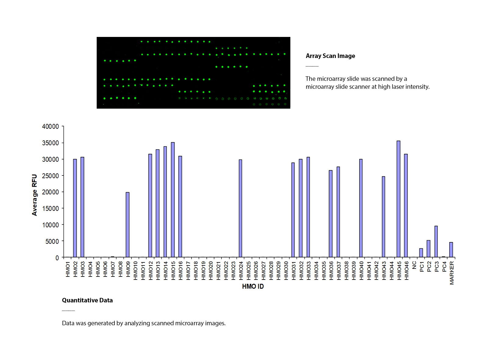
Aleuria Aurantia Lectin (AAL) binds to multiple human milk oligosaccharides
The HMO glycan array was assayed with biotinylated AAL lectin (10 μg/ml), followed by streptavidin-Cy3 (1 μg/ml). The array was scanned with a microarray scanner at 532nm wavelength. Positive control showed binding signals as expected. AAL binds to multiple human milk oligosaccharides.
List of human milk oligosaccharide structures on the array (download the PDF)
Protocol & User Manual (download the manual)