From: $994.00
Oligomannose glycan array technology was developed to allow researchers to explore the interactions between mannose-type glycans and various biological samples. It features 46 distinct oligomannose glycan structures, including linear, branched, high-mannose, and phosphorylated oligomannose glycans. These closely related isoforms with defined linkages offer a comprehensive group of glycans for studying mammalian and microbial mannose-binding proteins in details. This array system can be employed to analyze various biological samples (e.g. proteins, antibodies, cells, cell lysate, serum, vesicles, bacteria, or viral particles). The array system includes 8 or 16 identical subarrays, which enable simultaneous analysis of multiple samples. The assay format is simple, requires only a small sample volume, and provides reliable glycan-binding information with high throughput. The oligomannose glycan array can be customized to meet individual client needs, and assay services are available upon request.
Oligomannose glycans are present in various glycoproteins, and play critical roles in several biological processes. One essential function of oligomannoses is to facilitate the trafficking of lysosomal hydrolases by interacting with mannose-6-phosphate (Man-6-P) receptors, ensuring the correct delivery of lysosomal hydrolases to their destination. Additionally, oligomannoses attached to glycoproteins within the endoplasmic reticulum are necessary for proper protein folding and assembly. If a protein is misfolded, oligomannose glycans activate the protein quality control system, leading to protein degradation. This mechanism is crucial for maintaining cellular homeostasis and preventing the accumulation of misfolded or abnormal proteins, ensuring the proper functioning of cells.
In addition to their cellular functions, oligomannose glycans play a crucial role in immune recognition between hosts and microorganisms. Oligomannose glycans act as ligands for FimH, an adhesion protein located on the type 1 fimbriae of uropathogenic Escherichia coli, facilitating bacterial attachment and subsequent infection in human urinary epithelial cells. In contrast, oligomannose glycans act as pathogen-associated molecular patterns (PAMPs) in yeast and other microorganisms, and are recognized by a group of innate immune receptors known as C-type lectin receptors (CLRs), such as DC-SIGN, L-SIGN, Dectin-2, and Langerin. These CLRs are expressed on the surface of dendritic cells, macrophages, and other immune cells, and recognize oligomannose PAMPs to initiate immune responses. Due to their ability to interact with C-type lectin receptors on immune cells, oligomannose glycans play a critical role in shaping the innate immune response to microorganisms, providing host cells an essential defense mechanism against infections.
Although numerous oligomannose-binding proteins have been identified, their substrate specificity remains largely unexplored due to the lack of tools that facilitate relevant studies. To address this gap, Z Biotech has developed a robust oligomannose microarray platform that enables researchers to explore interactions between oligomannose glycans and various biological samples. The oligomannose array comprises 46 distinct oligomannose glycan structures, including linear, branched, high-mannose, and phosphorylated oligomannose glycans. These closely related isoforms with defined linkages offer a comprehensive group of glycans for studying mammalian and microbial mannose-binding proteins. The array system includes 8 or 16 identical subarrays, enabling the simultaneous analysis of multiple samples. The assay format is simple, requires only a small sample volume, and provides reliable glycan-binding information with high throughput.
Features
Applications
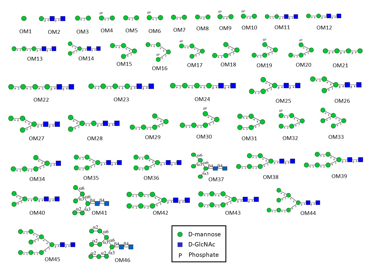
List of oligomannose structures on the array (download the PDF)
ID | Structure |
---|---|
OM1 | Man |
OM2 | Manβ1-4GlcNAcβ1-4GlcNAc |
OM3 | Manα1-2Man |
OM4 | Man6Pα1-2Man |
OM5 | Manα1-3Man |
OM6 | Man6Pα1-3Man |
OM7 | Manα1-4Man |
OM8 | Manα1-6Man |
OM9 | Man4Pα1-6Man |
OM10 | Man6Pα1-6Man |
OM11 | Manα1-3Manβ1-4GlcNAcβ1-4GlcNAc |
OM12 | Manα1-6Manβ1-4GlcNAcβ1-4GlcNAc |
OM13 | Manα1-2Manα1-3Manβ1-4GlcNAcβ1-4GlcNAc |
OM14 | Manα1-6(Manα1-3)Manβ1-4GlcNAcβ1-4GlcNAc |
OM15 | Manα1-6Manα1-6(Manα1-3)Man |
OM16 | Manα1-6Manα1-6(Man6Pα1-3)Man |
OM17 | Man6Pα1-6Manα1-6(Manα1-3)Man |
OM18 | Manα1-2Manα1-3(Manα1-6)Man |
OM19 | Manα1-2Manα1-3(Man6Pα1-6)Man |
OM20 | Manα1-2Manα1-3(Man4Pα1-6)Man |
OM21 | Manβ1-4Manβ1-4Manβ1-4Man |
OM22 | Manα1-2Manα1-2Manα1-3Manβ1-4GlcNAcβ1-4GlcNAc |
OM23 | Manα1-2Manα1-3Manα1-6Manβ1-4GlcNAcβ1-4GlcNAc |
OM24 | Manα1-2Manα1-6Manα1-6Manβ1-4GlcNAcβ1-4GlcNAc |
OM25 | Manα1-2Manα1-3(Manα1-6)Manβ1-4GlcNAcβ1-4GlcNAc |
OM26 | Manα1-3Manα1-6(Manα1-3)Manβ1-4GlcNAcβ1-4GlcNAc |
OM27 | Manα1-6Manα1-6(Manα1-3)Manβ1-4GlcNAcβ1-4GlcNAc |
OM28 | Manα1-3(Manα1-6)Manα1-6Manβ1-4GlcNAcβ1-4GlcNAc |
OM29 | Manα1-2Manα1-2Manα1-3(Manα1-6)Man |
OM30 | Manα1-2Manα1-2Manα1-3(Man6Pα1-6)Man |
OM31 | Manα1-2Manα1-3(Manα1-6Manα1-6)Man |
OM32 | Manα1-2Manα1-3(Man6Pα1-6Manα1-6)Man |
OM33 | Manα1-6(Manα1-3)Manα1-6(Manα1-3)Man |
OM34 | Manα1-2Manα1-2Manα1-3(Manα1-6)Manβ1-4GlcNAc |
OM35 | Manα1-2Manα1-3(Manα1-3Manα1-6)Manβ1-4GlcNAcβ1-4GlcNAc |
OM36 | Manα1-2Manα1-3(Manα1-6Manα1-6)Manβ1-4GlcNAcβ1-4GlcNAc |
OM37 | Manα1-6(Manα1-3)Manα1-6(Manα1-3)Manβ1-4GlcNAcβ1-4GlcNAc |
OM38 | Manα1-2Manα1-2Manα1-3(Manα1-3Manα1-6)Manβ1-4GlcNAcβ1-4GlcNAc |
OM39 | Manα1-2Manα1-2Manα1-3(Manα1-6Manα1-6)Manβ1-4GlcNAcβ1-4GlcNAc |
OM40 | Manα1-2Manα1-3(Manα1-2Manα1-6)Manα1-6Manβ1-4GlcNAcβ1-4GlcNAc |
OM41 | Manα1-6(Manα1-3)Manα1-6(Manα1-2Manα1-3)Manβ1-4GlcNAcβ1-4GlcNAc |
OM42 | Manα1-2Manα1-2Manα1-3(Manα1-2Manα1-3Manα1-6)Manβ1-4GlcNAcβ1-4GlcNAc |
OM43 | Manα1-2Manα1-2Manα1-3(Manα1-2Manα1-6Manα1-6)Manβ1-4GlcNAcβ1-4GlcNAc |
OM44 | Manα1-6(Manα1-3)Manα1-6(Manα1-2Manα1-2Manα1-3)Manβ1-4GlcNAcβ1-4GlcNAc |
OM45 | Manα1-2Manα1-6(Manα1-3)Manα1-6(Manα1-2Manα1-2Manα1-3)Manβ1-4GlcNAcβ1-4GlcNAc |
OM46 | Manα1-2Manα1-6(Manα1-2Manα1-3)Manα1-6(Manα1-2Manα1-2Manα1-3)Manβ1-4GlcNAcβ1-4GlcNAc |
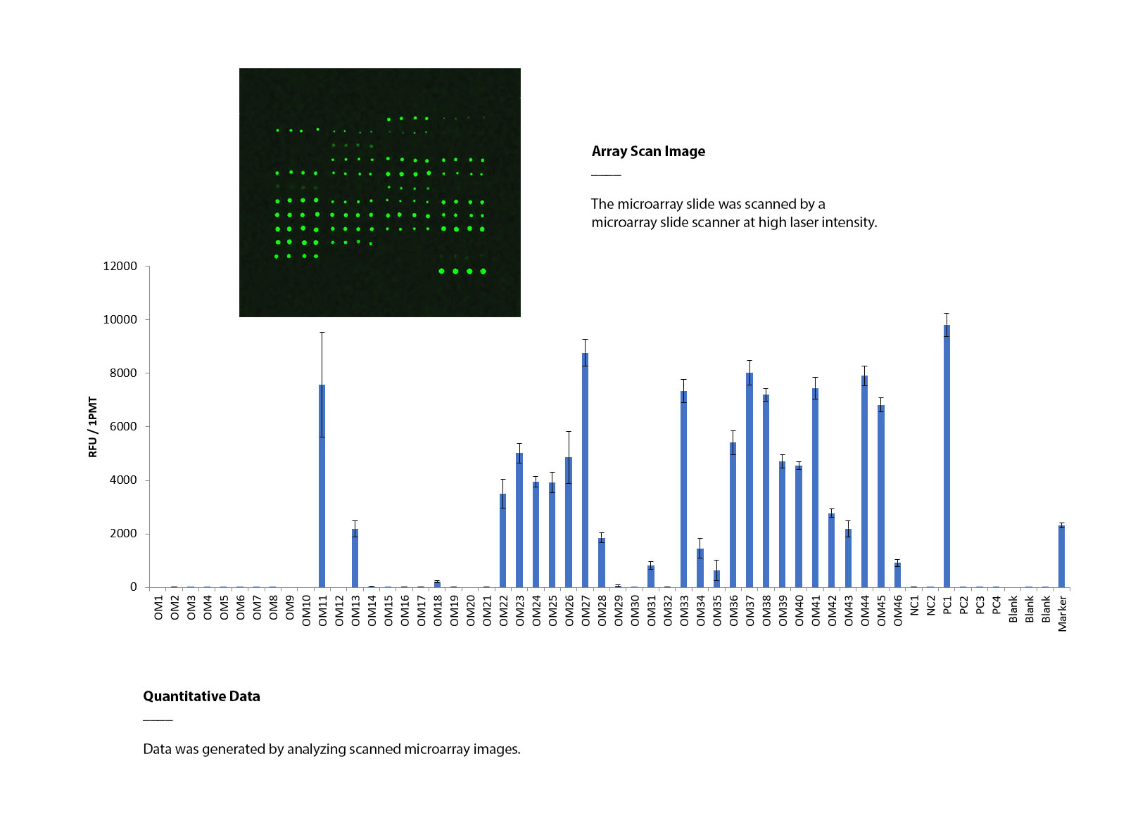
Using oligomannose glycan array to determine the binding specificity of Galanthus nivalis lectin (GNA)
The oligomannose glycan array was probed with biotinylated Galanthus nivalis lectin (GNA) at a concentration of 10 μg/mL, followed by streptavidin labeled with Cy3. The array was scanned using a microarray scanner at a wavelength of 532 nm. The positive control (PC1) exhibited expected binding signals, and GNA demonstrated differential interactions with various oligomannose glycans. These results provide evidence of the array’s functionality in probing oligomannose-binding proteins and studying their interactions with oligomannose glycans.
List of oligomannose structures on the array (download the PDF)
Protocol & User Manual (download the manual)