Pan-Sialylation Detection Kit

10901-30T

From: $399.00

The Pan-Sialylation Detection Kit enables researchers to analyze the sialylation (presence of sialic acids) of mammalian live cells by flow cytometry. This innovative method uses mild periodate oxidation to generate an aldehyde on sialic acids, followed by aniline-catalyzed oxime ligation with biotin tags. The ligated biotin is then detected by fluorescently labeled streptavidin for flow cytometry analysis. Unlike lectin-based methods, which typically only detect alpha 2,3/2,6 sialic acids on specific glycans, this kit detects pan-sialic acids on a broader range of glycans. The assay kit includes all necessary reagents for up to 30 tests, along with a detailed protocol to guide researchers step-by-step to achieve optimal results. In addition to the assay kit, we offer related services to perform the analysis, tailored to meet your specific experimental needs.

SKU: 10901-30T Category:
Description
Examples

Description

Sialic acid (Neu5Ac) is a family of nine-carbon sugars typically found at the terminal positions of glycan chains on glycoproteins and glycolipids on the surface of cells. Sialylation refers to the process of adding sialic acid residues to these glycan chains. This modification plays a crucial role in a variety of biological processes, including cell-cell interactions, immune response regulation, and pathogen recognition. Aberrant sialylation is associated with numerous diseases, including cancer, inflammation, and infectious diseases, making it a critical area of study for understanding and diagnosing various health conditions.

The Method Behind Pan-Sialylation Detection

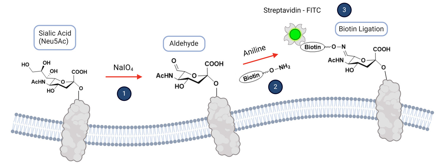
The Pan-Sialylation Detection Kit utilizes a robust and detailed chemical method to detect sialic acids on the surface of live cells via flow cytometry (Figure 1):

  1. Mild Periodate Oxidation:

The first step involves the mild oxidation of sialic acids on the cell surface using sodium periodate. This reagent specifically targets the vicinal diol groups present in sialic acids, oxidizing them to form reactive aldehyde groups. The oxidation is mild enough to preserve cell integrity, ensuring that the cells remain viable for subsequent analysis.

  1. Aniline-Catalyzed Oxime Ligation:

Next, the generated aldehyde groups on the sialic acids are subjected to aniline-catalyzed oxime ligation with aminooxy-biotin. Aniline acts as a nucleophilic catalyst, enhancing the formation of the oxime bond between the aldehyde groups and the aminooxy-biotin. Aniline facilitates the nucleophilic attack of the aminooxy group on the aldehyde, forming a stable oxime linkage and effectively tagging the sialic acids with biotin.

  1. Detection with FITC-Conjugated Streptavidin:

Finally, the biotinylated sialic acids are detected using FITC-conjugated streptavidin. Streptavidin binds strongly to biotin, and the fluorescent FITC label allows for the detection of these complexes via flow cytometry. The fluorescence intensity correlates with the amount of sialylation present on the cell surface, providing a quantitative measure of sialic acid levels.

Figure 1. Illustration of Sialic Acid Labeling with Biotin for Flow Cytometry Analysis

Assay Kit Specifications

The Pan-Sialylation Detection Kit includes all the necessary reagents for performing up to 30 tests. Each kit contains:

  • Sodium periodate for mild oxidation
  • Aminooxy-biotin for ligation
  • FITC-conjugated streptavidin for detection
  • Ligation buffer containing aniline
  • Additional buffers required for the assay
  • Detailed protocol

For optimal results, we recommend using 2.0-5.0 x 10^5 cells per analysis. This cell range ensures sufficient detection and accurate analysis of sialylation levels. To determine the optimal concentration of sodium periodate for different cell types, we suggest performing a concentration optimization experiment. This involves testing 3-5 recommended concentrations of sodium periodate to oxidize the cells and determine the saturation point. This step is crucial since the level of sialylation can vary among different cell types, and identifying the best concentration will maximize the efficiency and accuracy of the assay.

Customization and Services

In addition to the assay kit, we offer customized assay services tailored to meet specific experimental requirements, allowing researchers to leverage our expertise for their unique research needs.

Examples

Oxidation Levels and Sialic Acid Labeling

Human melanoma cell line A375 cells were oxidized with sodium periodate at various concentrations to achieve different levels of oxidation. Following oxidation, the cells were ligated with biotin using the same protocol and analyzed for sialic acid labeling. The results indicate that varying levels of oxidation by sodium periodate correlate with the degree of sialic acid labeling with biotin tags (Figure 1).


Figure 1. Concentrations of sodium periodate affect the oxidation levels, which directly correlate with the levels of biotin labeling on the sialic acids.

Neuraminidase Treatment and Sialic Acid Detection

A375 cells were treated with either a control solution or neuraminidase from Clostridium perfringens (400 mU/mL). The cells were then analyzed using the Pan-Sialylation Detection Kit, with sodium periodate used at a concentration of 0.5 mM as indicated in Figure 2. The data demonstrate that neuraminidase treatment significantly reduces the amount of sialic acid present on the cell surface, reflected by the decreased levels of biotin-labeled sialic acids.


Figure 2. Neuraminidase treatment significantly reduces the presence of sialic acids on the cell surface, as reflected by the reduced levels of biotin-labeled sialic acids.

Validation with Sialic Acid-Recognizing Lectins
To validate the results indicated by the biotin-labeled sialic acids, control and neuraminidase-treated cells were analyzed using sialic acid-recognizing lectins SNA and MALII. The data show that neuraminidase treatment reduces the presence of sialic acids on the cell surface, consistent with the findings using the Pan-Sialylation Detection Kit.


Figure 3. Neuraminidase treatment reduces SNA and MALII binding, confirming the reduction of sialic acids on the cell surface.