SARS-CoV-2 RBD Peptide and Glycopeptide Array

10801

From: $360.00

The SARS-CoV-2 receptor-binding domain (RBD) peptide and glycopeptide array allows researchers to identify, profile, and compare specific IgG, IgA, and IgM antibody responses or detect and validate protein interactions with various RBD peptides and glycopeptides in a customizable format. Researchers can choose from a list of 83 wild-type and mutant RBD peptides and glycopeptides (15-mer in length), including sequences from the Alpha, Beta, Gamma, Delta, Omicron, and other SARS-CoV-2 variants. The selected peptides/glycopeptides are immobilized on a solid microarray surface for high-throughput binding analysis.

SKU: 10801 Category:
Description
Structures
Examples
Document

Description

Coronavirus Disease 2019 (COVID-19), an infectious disease resulting from the Severe Acute Respiratory Syndrome Coronavirus 2 (SARS-CoV-2) infection, has ravaged the world since December 2019. Throughout history, there have been several coronavirus outbreaks, including Severe Acute Respiratory Syndrome (SARS) during 2002-2003 in South East Asia and Middle East Respiratory Syndrome (MERS) in 2012 in the Middle East and 2015 in Korea. However, none of these outbreaks caused such a significant impact on public health as COVID-19.

Coronavirus enters the host cells by either host receptor-mediated fusion or fusing after being swallowed into an endosome.  For example, the spike (S) protein of SARS-CoV-2 plays a vital role in receptor recognition and the cell membrane fusion process.  The spike (S) protein comprises two subunits, S1 and S2.  The S1 subunit contains a receptor-binding domain (RBD) that recognizes and binds to the host receptor, while the S2 subunit mediates viral cell membrane fusion.  For example, SARS-CoV-2 viruses utilize the human angiotensin-converting enzyme 2 (ACE2) type I membrane protein as an entry receptor. Upon binding to the ACE2 receptor by viral S protein, with the help of transmembrane serine protease 2 (TMPRSS2) on the cell surface, SARS-CoV-2 enters the host cells through direct membrane fusion. Therefore, identifying and characterizing host RBD-binding protein will accelerate our understanding of the virus and potentially unveil new targets for therapeutic interventions. As the humoral immune response is a central part of SARS-CoV-2 – elicited immunity, uncovering potent RBD-neutralizing antibodies is essential for developing new modalities to tackle the virus.

Z Biotech has developed a robust microarray platform that allows researchers to identify, profile, and compare specific IgG, IgA, and IgM antibody responses or detect and validate protein interactions with various RBD peptides and glycopeptides in a customizable format. Researchers can choose from a list of 83 wild-type and mutant RBD peptides and glycopeptides (15-mer in length), including sequences from the Alpha, Beta, Gamma, Delta, Omicron, and other SARS-CoV-2 variants. The selected peptides/glycopeptides are immobilized on a solid microarray surface for high-throughput binding analysis.

Features

  • Unrivaled sensitivity and specificity;
  • Simple assay format;
  • Small sample volume;
  • Customizable (select peptides and glycopeptides for a specific microarray format)
  • Assay service available;

Applications

  • Profile antibody responses from the patient or animal sera;
  • Screen and characterize immunodominant RBD epitopes;
  • Compare antibody responses across various RBD mutants.
  • Identify RBD-interacting proteins;

Structures

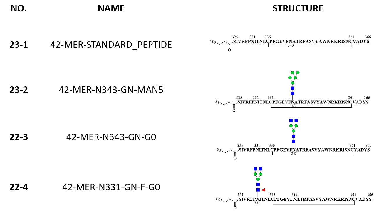
List of peptides and glycopeptides for custom RBD microarray (download the PDF)

List of peptides for custom RBD microarray; additional peptides available upon request.

Controls:  SARS-CoV-2 S Trimer protein, S1 subunit protein, S2 subunit protein, N protein, BSA, hACE2, streptavidin-Cy3, streptavidin-Cy5, Mouse IgM, Mouse IgG, Rabbit IgM, Rabbit IgG, Human IgM, Human IgG, GST, His-biotin.

No. Sequence Type Start End
1 RVQPTESIVRFPNIT WT 319 333
2 FPNITNLCPFGEVFN WT 329 343
 2-a FPNITNLCPFDEVFN Mutant
3 GEVFNATRFASVYAW WT 339 353
 3-a GEVFKATRFASVYAW Mutant
 3-b GEVFNATIFASVYAW Mutant
 3-c GEVFNATKFASVYAW Mutant
 3-d GEVFNATTFASVYAW Mutant
4 SVYAWNRKRISNCVA WT 349 363
4-a SVYAWKRKRISNCVA Mutant
5 SNCVADYSVLYNSAS WT 359 373
5-a SNCVADYSVLYNLAP Mutant
6 YNSASFSTFKCYGVS WT 369 383
6-a YNLAPFFTFKCYGVS Mutant
7 CYGVSPTKLNDLCFT WT 379 393
7-a CYGLSPTKLNDLCFT Mutant
7-b CYGVSLTKLNDLCFT Mutant
8 DLCFTNVYADSFVIR WT 389 403
9 SFVIRGDEVRQIAPG WT 399 413
9-a SFVIRGDEVRQMAPG Mutant
10 QIAPGQTGKIADYNY WT 409 423
10-a QIAPGQTGNIADYNY Mutant
10-b QIAPGQTGTIADYNY Mutant
10-c QIAPGQTGMIADYNY Mutant
10-d QIAPGQTGRIADYNY Mutant
11 ADYNYKLPDDFTGCV WT 419 433
12 FTGCVIAWNSNNLDS WT 429 443
12-a FTGCVVAWNSNNLDS Mutant
12-b FTGCVIAWNSKNLDS Mutant
12-c FTGCVIAWNSNKLDS Mutant
13 NNLDSKVGGNYNYLY WT 439 453
13-a NKLDSKVSGNYNYLY Mutant
13-b NNLDSKVDGNYNYLY Mutant
13-c NNLDSKVVGNYNYLY Mutant
13-d NNLDSKVAGNYNYLY Mutant
13-e NNLDSKVGGNFNYLY Mutant
13-f NNLDSKVGGNCNYLY Mutant
13-g NNLDSKVGGNSNYLY Mutant
13-h NNLDSKVGGNYNYRY Mutant
13-i NNLDSKVGGNYNYQY Mutant
14 YNYLYRLFRKSNLKP WT 449 463
14-a YNYRYRLFRKSNLKP Mutant
14-b YNYPYRLFRKSNLKP Mutant
14-c YNYQYRLFRKSNLKP Mutant
14-d YNYLYRFFRKSNLKP Mutant
14-e YNYLFRLFRKSNLKP Mutant
14-f YNYLSRLFRKSNLKP Mutant
15 SNLKPFERDISTEIY WT 459 473
16 STEIYQAGSTPCNGV WT 469 483
16-a STEIYQVGSTPCNGV Mutant
16-b STEIYQGGSTPCNGV Mutant
16-c STEIYQAGNTPCNGV Mutant
16-d STEIYQAGTTPCNGV Mutant
16-e STEIYQAGITPCNGV Mutant
16-f STEIYQAGRTPCNGV Mutant
16-g STEIYQAGSKPCNGV Mutant
16-h STEIYQAGSRPCNGV Mutant
16-i STEIYQAGSIPCNGV Mutant
17 PCNGVEGFNCYFPLQ WT 479 493
17-a PCNGVKGFNCYFPLQ Mutant
17-b PCNGVQGFNCYFPLQ Mutant
17-c PCNGVAGFNCYFPLQ Mutant
17-d PCNGVEGFNCYSPLQ Mutant
17-e PCNGVEGFNCYYPLQ Mutant
18 YFPLQSYGFQPTNGV WT 489 503
18-a YSPLQSYGFQPTNGV Mutant
18-b  YFPLRSYSFRPTYGV Mutant
18-c YFPLQAYGFQPTNGV Mutant
18-d YFPLQPYGFQPTNGV Mutant
18-e YFPLQTYGFQPTNGV Mutant
18-f YFPLQSYGFQPTYGV Mutant
18-g YFPLQSYGFQPTRGV Mutant
18-h YFPLQSYGFQPTDGV Mutant
18-i YFPLQSYGFQPTHGV Mutant
19 PTNGVGYQPYRVVVL WT 499 513
19-1 PTYGVGYQPYRVVVL Mutant
19-2 PTYGVGHQPYRVVVL Mutant
20 RVVVLSFELLHAPAT WT 509 523
20-a RVVVLSFELLHTPAT Mutant
20-b RVVVLSFELLHSPAT Mutant
20-c RVVVLSFELLHPPAT Mutant
21 HAPATVCGPKKSTNL WT 519 533
22 KSTNLVKNKCVNF WT 529 541

List of glycopeptides for custom RBD microarray; additional glycopeptides available upon request.

Examples

SARS-CoV-2 spike RBD antibodies differentially interact with RBD peptides

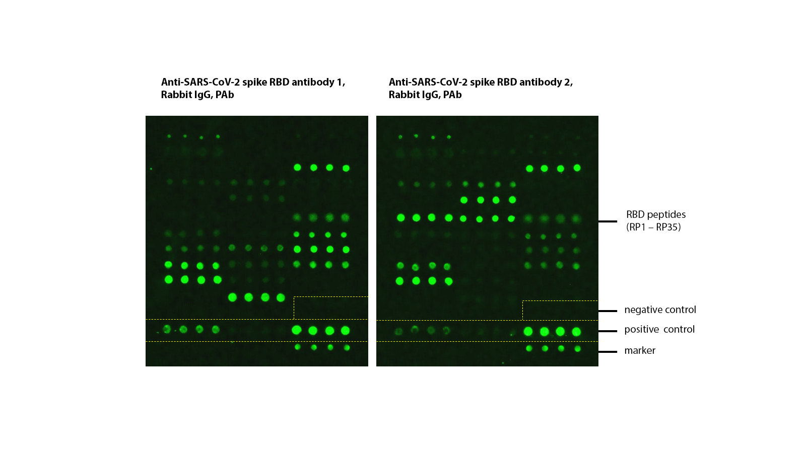
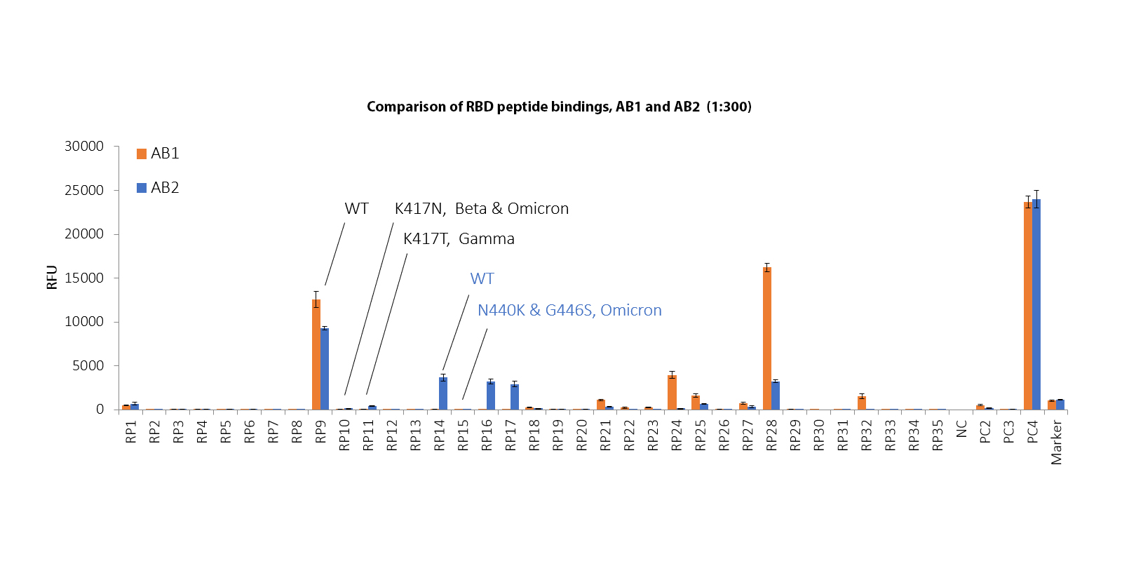
35 RBD peptides, either WT or mutant, were selected to create a custom microarray. This RBD microarray was assayed with two commercially available SARS-CoV-2 (2019-nCoV) spike RBD antibodies (rabbit PAb, 1:300), followed by a secondary antibody (anti-rabbit-IgG, Cy3). The array was scanned with a microarray scanner at 532nm wavelength. Positive control 3 (PC3, rabbit IgG) showed binding signals as expected, as well as multiple RBD peptides. Negative control (NC) showed no binding signal.

Both AB1 and AB2 bind to WT RBD peptide 9 (PR9). For the same peptide, the bindings to K417N (PR10) and K417T (PR11) mutants are lost. AB2 binds with WT peptide 14 (RP14) while completely losing its binding capacity to the Omicron mutant (RP15).

Document

List of peptides and glycopeptides for custom RBD microarray (download the PDF)

Protocol & User Manual (download the manual)